This report details the clinical case of a 54-year-old female patient who presented with a sudden loss of vision in her right eye, accompanied by severe headaches. The patient described the onset of her symptoms as feeling like a "curtain" had been drawn over her vision. Upon arrival at the clinic, she was visibly nervous and tense, having already explored numerous unsuccessful treatment avenues.
The patient’s medical history was unremarkable, with no prior surgeries or chronic diseases. Comprehensive hospital examinations, including MRI and CT scans, returned negative results. There was no history of trauma, and markers for arterial inflammation were also negative. Because standard clinical testing failed to establish a specific etiology, the condition was diagnosed by Dr. Larry Wallace as ischemic optic neuropathy or optic neuritis, potentially triggered by a stress response.
Prior to seeking coMra therapy, the patient had been treated at a hospital with high-dose prednisone (steroids) and underwent extensive diagnostic testing, yet her condition remained unchanged.
At the start of care, the patient’s right eye visual acuity was measured at 20/50-3. Her near vision was significantly compromised, showing reductions in both contrast sensitivity and acuity. Furthermore, her visual fields were restricted, characterized by enlarged blind spots and a reduced color field.
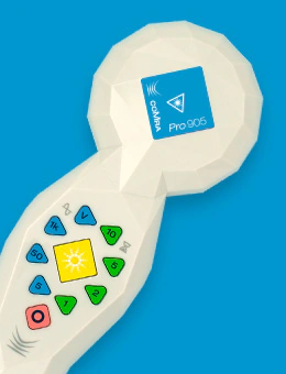
The treatment plan consisted of 18 sessions conducted over a 10-week period. The primary protocol utilized was Neurology 3, which was modified to include a scanning technique along the optic nerve pathway, moving along the side of the face and over the eye. The coMra Delta (specifically the 905 nm laser model) was used for these applications. This was supplemented by weekly acupuncture sessions.
Following the 18-treatment course, follow-up examinations revealed significant improvements: * Visual Acuity: Improved by approximately 20% for both near and far vision. * Contrast Sensitivity: Improved by nearly 30%, indicating a better ability to distinguish between light and dark. * Visual Fields: The patient’s blind spots were reduced, color fields expanded, and overall visual field defects decreased.
At the start of care, the patient presented with 14 distinct visual field defects. By the end of the treatment period, these had been reduced to nine, with a marked decrease in the severity of the remaining defects; notably, there were no longer any "absolute" defects. Throughout this period, the patient was successfully tapered off prednisone. Subjectively, she reported clearer vision at both distances, improved peripheral vision, and a significant reduction in the psychological disturbance caused by her field loss.
A follow-up assessment conducted one month later showed continued improvement in visual field defects, which further decreased from nine to five. While there was a minor fluctuation in acuity, the patient’s subjective experience remained positive, with continued clarity and improved peripheral awareness.
This case report highlights the potential of coMra therapy in supporting visual rehabilitation. A special acknowledgment is extended to Dr. Larry Wallace, a behavioral optometrist with over 40 years of experience and a key figure in the College of Syntonic Optometry. Dr. Wallace is a certified low-vision specialist dedicated to research in light and color therapy, particularly regarding visual rehabilitation for head trauma and brain injury. As the patent holder for the first microcurrent device used to treat retinal disease, Dr. Wallace continues to travel globally to educate practitioners on the applications of syntonic phototherapy.