

Why Some Optic Nerves Heal – and Others Don’t
The mitochondria decide – powerhouse or alarm system?
"The optic nerve damage is permanent. We can't restore what's lost. We'll manage the pressure and monitor for progression, but the vision that's gone isn't coming back."
Most clinicians have delivered this news many times.
But what if...
Severe, complete nerve death with scarring is irreversible. But what about the patients who don't fit that picture? The ones with damaged but not dead tissue? The ones still in that critical window where the right support might shift the outcome?
What if peer-reviewed, reproducible clinical research shows that measurable functional recovery is possible in the exact types of patients being told "there's nothing more we can do"?
Not fringe science. Not anecdotal miracles. Published research with objective clinical endpoints.
134 patients with primary open-angle glaucoma—early, moderate, and advanced stages.
Advanced-stage patients experienced 74.4% visual field expansion. Scotomas disappeared in 58.2% of cases.
Visual evoked potentials increased by 41.4%, indicating improved neural function from retina to brain.
27 patients with optic nerve atrophy from various causes saw 65% improvement in visual acuity on average—from near-blindness (0.07) to functional vision (0.2).
Twelve moved from below 0.1 to 0.1 or higher. Absolute scotomas decreased by 35.4%.
These are not marginal changes. They represent functional recovery in conditions conventionally described as irreversible.
Which begs the question: is it a biological fact that "the damage is permanent"?
The Mechanism Conventional Training Overlooked
Consider a glaucoma patient whose vision keeps deteriorating despite perfect IOP control: their mitochondria—the powerhouses that fuel every cellular process in those energy-hungry retinal ganglion cells—are stuck in alarm mode.
In glaucoma and other optic neuropathies, retinal ganglion cells depend on dense mitochondrial networks to sustain vision.
But under sustained stress, mitochondria don't simply fail to make ATP—they change identity.
They flip from powerhouses into alarm systems.
When that happens, mitochondria leak fragments of mitochondrial DNA, reactive oxygen species, cytochrome c, and metabolites such as succinate and fumarate into the surrounding environment.
These are known as DAMPs—danger-associated molecular patterns. They do exactly what the name suggests: they signal danger.
A deteriorating patient's tissue may well be screaming for help.
These DAMPs activate innate immune pathways, triggering inflammasomes and cytokine cascades that lock tissue into chronic inflammation. The tissue shifts from "repair this damage" to "this area is under attack—defend and destroy."
Lowering intraocular pressure helps, but it doesn't turn off these cellular alarms. It doesn't stop the DAMP leak. It doesn't halt the inflammatory lock-in that's driving progressive neurodegeneration.
That is why a patient's visual field may continue to shrink despite perfect IOP control: the mitochondrial crisis persists, unseen and untreated.
When the Body Knows Better
Western medicine operates on a control paradigm: identify the problem, apply the correction, override the symptom.
But the body's healing intelligence often works in ways that contradict our training.
Take shock, for example. When the body experiences severe trauma, it actively down-regulates almost every process—blood shunts to core organs, metabolism slows, inflammatory responses dampen. If the body responded proportionally to the insult, you would die. Full inflammatory cascade, maximum metabolic demand, complete system activation—these "logical" responses would be fatal.
Instead, the body does something counterintuitive: it goes quiet. It enters a protective state that medical science still doesn't fully understand—but that consistently saves lives.
The body possesses survival intelligence we don't have.
When some patients stabilize while others progress despite identical treatment, the body is making decisions based on tissue state, systemic reserves, and healing capacity that we can't fully measure.
Our role isn't to override that intelligence. It's to support it.
The Fork in the Road: Sub-Lethal Mitochondrial Stress
Not every damaged cell dies immediately. There's a middle ground—a state researchers call sub-lethal mitochondrial stress.
In this state:
- Cells remain alive but dysfunctional
- Mitochondria leak moderate amounts of danger signals without triggering full apoptosis
- Inflammatory signaling becomes chronic rather than resolving
- The tissue is locked in a slow burn toward progressive degeneration
This is the window of opportunity—the moment when support for mitochondrial stability can determine whether tissue recovers or declines.
Monitoring does not fix mitochondrial dysfunction.
But supporting those struggling mitochondria—helping them shift from alarm mode back to stable function—can change outcomes dramatically.
Evidence of Functional Recovery
Study 1: Glaucoma Recovery (134 Patients)
Combined transcranial magnetic and infrared laser therapy versus standard pressure management alone.
Results:
- Visual field expansion for white light: 56.4% on average
- Advanced-stage patients: 74.4% expansion
- Scotomas completely disappeared: 58.2% of cases (versus 22.5% in pressure-only controls)
- Visual evoked potential amplitude: +41.4% (objective measure of retina-to-brain signal strength)
- Optic nerve blood flow: 19.3% improvement
These were measured improvements on perimetry, VEP testing, and Doppler imaging.
The control group received excellent IOP management—the standard of care. They continued to decline.
The difference wasn't better pressure control. It was mitochondrial support.
Study 2: Optic Nerve Atrophy (27 Patients)
Patients received a single session of combined electrical and laser stimulation directly to the optic nerve (invasive procedure).
Results measured over months:
- Visual acuity improved 65% on average (from 0.07 to 0.2)
- 12 patients (44%) went from near-blindness (<0.1) to functional vision (≥0.1)
- Absolute scotomas reduced by 35.4%
- Foveal light sensitivity increased by 22.3%
- Visual fields expanded by 10-20 degrees in responders
Histological examination showed structural reorganization of myelin sheaths around surviving axons. Measurable tissue-level repair.
The researchers documented increased nerve conduction velocity and enhanced membrane excitability—objective markers of functional recovery in tissue usually diagnosed as permanently damaged.
Study 3: Neurotrophic Factors (217 Patients)
A follow-up investigation measured neurotrophic factors—proteins like brain-derived neurotrophic factor (BDNF) and ciliary neurotrophic factor (CNTF) that support neuronal survival—in 217 glaucoma patients.
One group received magnetic-laser therapy in addition to standard IOP management. The control group received IOP management alone.
Both groups achieved target IOP. Both had perfect pressure control.
Over one year:
- Treatment group: stabilization or improvement in visual function
- Control group: continued deterioration
The difference? Neurotrophic factor levels.
BDNF increased significantly in the treatment group. It showed strong correlation (r = 0.81) with retinal ganglion cell activity on electrophysiology testing.
Translation: Even with perfect pressure control, patients decline because the cellular mechanisms that determine whether neurons survive or die are not addressed.
The Missing Tools
This gap exists because medical training rarely covers mitochondrial therapeutics.
There are few approved options designed to:
- Enhance ATP production in damaged neural tissue
- Reduce mitochondrial danger signaling
- Support neurotrophic factor expression
- Modulate inflammatory lock-in that drives neurodegeneration
CoQ10 and NAD+ precursors show promise in research, but they're not part of standard ophthalmic protocols. Idebenone exists for one rare condition in Europe.
That's the extent of the mitochondrial toolkit most clinicians were given.
Research demonstrating functional recovery has focused on biophysical approaches—combined magnetic fields, infrared light, and in some cases ultrasound—that influence cellular energetics through mechanisms drugs can't replicate.
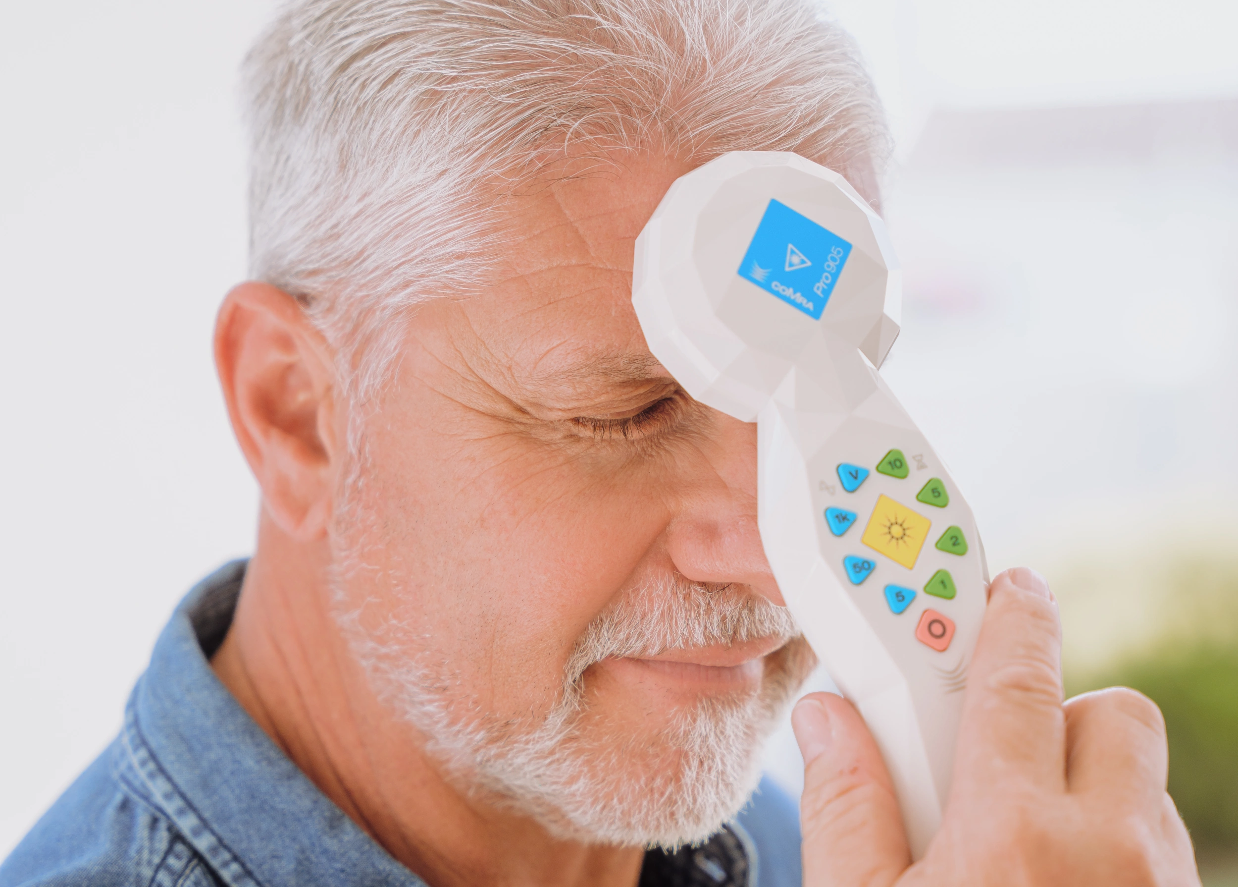
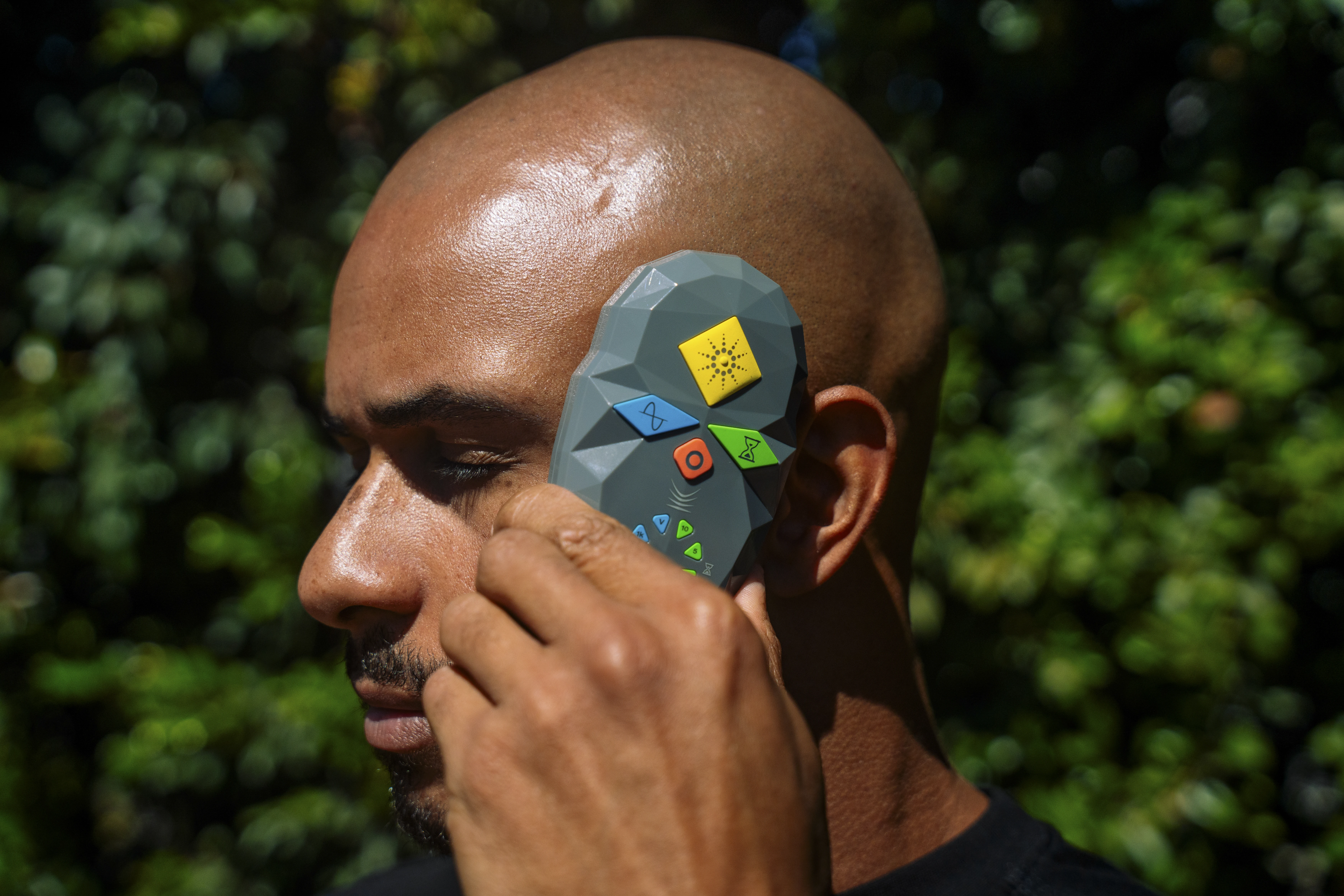
Among these approaches, coMra therapy combines, near-infrared laser, magnetic fields, ultrasound, and color modulation to support mitochondrial coherence and resilience.
The critical distinction: these approaches provide signals, not substances.
Light, magnetic fields, and ultrasound don't force biochemical pathways—they create conditions for coherence. They work with the body's intelligence rather than overriding it. The body has a rhythm. coMra therapy is designed to support that rhythm, not override it.
Research on biophysical modalities documents:
- 200-300% increases in ATP production through photomagnetic catalysis
- Improved neurotrophic factor expression
- Phase-specific modulation that matches tissue needs
- Measurable clinical outcomes in conditions conventionally called irreversible
These modalities exist. The research exists. But they weren't in most textbooks.
Understanding why outcomes vary with these approaches—and what that reveals about the body's healing process—is critical to expanding clinical options responsibly.
Why Outcomes Vary: Only the Body Can Heal the Body
If you explore approaches like coMra therapy that support mitochondrial function, you'll encounter something uncomfortable: outcomes can vary significantly.
Some patients with advanced glaucoma experience 74% visual field expansion. Others stabilize. Some show minimal response.
Why the variability?
Because only the body can heal the body.
Biophysical approaches that support mitochondrial coherence don't force a biochemical outcome—they engage the body's innate intelligence to guide the healing process.
If viable nerve tissue remains, if the inflammatory cascade can be interrupted, if mitochondria can shift from alarm mode to stable function—recovery becomes possible. But the body determines what's possible based on that specific tissue state.
The approach isn't about power—it's about signal. Not about forcing—it's about supporting. Not about overriding the body's intelligence—it's about cooperating with it.
When research shows 56% average visual field expansion in glaucoma, that's not a promise that every patient will achieve that result. It's evidence that when mitochondrial function is supported, functional recovery becomes possible—and the body's healing intelligence determines the extent.
Some patients experience dramatic recovery. Others stabilize—vision stops declining, which for progressive disease is a significant win. Others slow their decline when complete recovery isn't possible.
The body makes that determination based on:
- How much viable tissue remains versus scar tissue
- Systemic reserves and overall health status
- The duration and severity of the mitochondrial crisis
- Individual healing capacity we can't fully measure
We provide support. The body determines the outcome.
The Deeper Question
How many patients in active care have viable but declining tissue—cells in that sub-lethal window that could still recover if their mitochondria received proper support?
This is not a question of blame, but of awareness.
Every profession evolves by revisiting assumptions once thought settled.
If functional recovery has been documented and is reproducible, then remaining curious is not optional—it's part of clinical responsibility.
Expanding the Toolkit
Standard ophthalmic care—IOP management, pharmacology, surgery—remains essential.
But if the deciding factor in nerve survival is mitochondrial stability, then expanding the toolkit to include modalities that restore it is both logical and ethical.
Approaches such as coMra therapy add this missing dimension.
They don't replace standard care—they complete it.
This is not about guilt. It's about growth.
About giving every patient the best possible chance at preserving sight.
What Comes Next
The evidence is clear: functional recovery in optic nerve disease is possible when mitochondrial function is supported appropriately.
The next step is understanding how to apply that knowledge—how biophysical signals influence mitochondria, how coherence supports repair, and how clinical protocols can align with the body's own rhythm of healing.
If this raises new questions—that's the point.
Progress begins with discomfort, followed by curiosity, then exploration.
How does the damage progress—and what stops it?
Subscribe below to receive our next article on mitochondrial alarm signals and clinical intervention.
References
Egorov E.A., Kamenskikh T.G., Raigorodsky Yu.M., Kolbenev I.O., Kamenskikh I.D. (2013). The results of transcranial application of the low-intensity magneto-laser treatment in the patients presenting with primary open-angle glaucoma. _Fizioterapiya, Balneologiya i Reabilitatsiya_, 5, 15-18.
Gadzhieva N.S., Linnik L.F., Rudneva M.A. (1994). Method of one-time combined electrical and laser stimulation of the optic nerve in the treatment of atrophy of various genesis. _Ophthalmosurgery_, 1, 47-54.
Egorov E.A., Kamenskikh I.D., Kamenskikh T.G., Kolbeneva I.O., Raygorodsky Yu.M., Tushin V.V. (2016). Influence of dynamic transcranial magnetotherapy and laser stimulation on the activity of retinal ganglion cells and the level of neurotrophic factors. _RMJ Clinical Ophthalmology_, 1, 19-24.